Home » Without Label » Shoulder Anatomy Diagram : Shoulder Anatomy Sketch Scapula And Humerus Bone By Vectortradition / Starting with what is deepest, it goes:
Shoulder Anatomy Diagram : Shoulder Anatomy Sketch Scapula And Humerus Bone By Vectortradition / Starting with what is deepest, it goes:
Shoulder Anatomy Diagram : Shoulder Anatomy Sketch Scapula And Humerus Bone By Vectortradition / Starting with what is deepest, it goes:. The anterior shoulder pain usually develops when injury or inflammation occurs in the tendons that are attached to the shoulder joint. There are many nerves and blood vessels that supply the muscles and bones of the shoulder. Other important bones in the shoulder include: The shoulder is a complex combination of bones and joints where many muscles act to provide the widest range of motion of any part of the body. However, more serious injuries, such as complete rotator cuff tears, may require surgical repair.
Four of them are found on the anterior aspect of the shoulder, whereas the rest are located on the shoulder's posterior aspect and in the back. All of the nerves that travel down the arm pass through the axilla (the armpit) just under the shoulder joint and are known as the brachial plexus before dividing into the individual nerves.these nerves carry the signals from the brain to the muscles that move the arm. Pulls rhomboideus major raises and adducts the scapula. The glenohumeral joint is where the ball (humeral head) and the socket (the glenoid) meet. The shoulder joint (glenohumeral joint) is a ball and socket joint between the scapula and the humerus.it is the major joint connecting the upper limb to the trunk.
Shoulder Anatomy In Detail from www.anatomynote.com The different types of connective tissues in the shoulder are bone articular cartilage ligaments joint capsules and bursa see gross anatomy. This diagram depicts shoulder anatomy muscles diagram.human anatomy diagrams show internal organs, cells, systems, conditions, symptoms and sickness information and/or tips for healthy living. Deltoides triangular refers to the front head of the. The shoulder joint is the junction between the chest and the upper extremity. The shoulder joint is the connection between the chest and the upper extremity. Normal anatomy, variants and checklist. Two joints are at the shoulder. Anatomy of the shoulder part 3 (muscular structures).
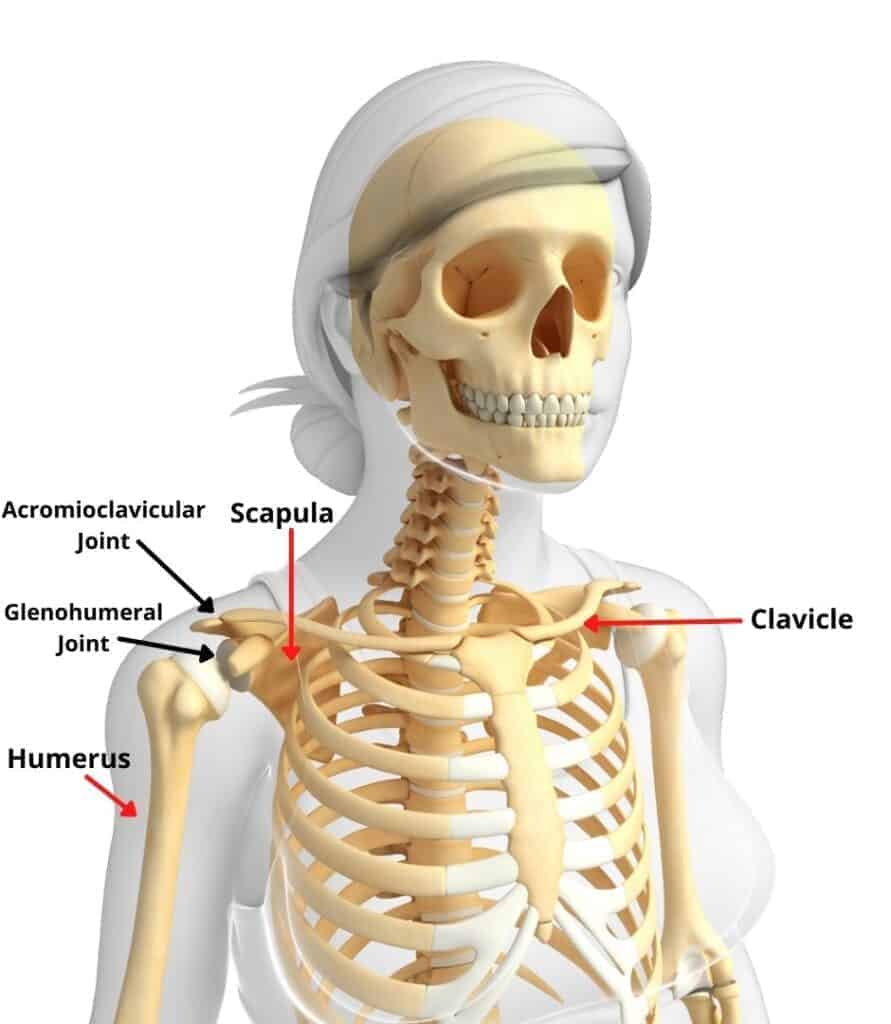
The large bone in the upper arm is called the humerus.
The shoulder is made up of two joints, the acromioclavicular joint and the glenohumeral joint. The top of the humerus is shaped like a ball. This diagram depicts shoulder anatomy muscles diagram.human anatomy diagrams show internal organs, cells, systems, conditions, symptoms and sickness information and/or tips for healthy living. There are many nerves and blood vessels that supply the muscles and bones of the shoulder. The shoulder joint is formed where the humerus upper arm bone fits into the scapula shoulder blade like a ball and socket. Bone, then ligaments of the joint capsule, with tendons and muscles on top. It causes pain in the area just outside the joint. A second joint in the shoulder is the junction of the collar bone with the shoulder blade, called the. Diagram of human forearm bones anatomy 10 photos of the diagram of human forearm bones anatomy anatomy elbow bones, anatomy shoulder bones, hand bones anatomy, human anatomy arm bones, leg bones anatomy, two bones in the forearm, wrist bones anatomy, hand, anatomy elbow bones, anatomy shoulder bones. Male shoulder ligaments and biceps muscles isolated in skeleton labeled chart on white labeled human anatomy diagram of male shoulder ligaments, connective tissue and biceps muscles isolated within the skeletal system frontal anterior view on a white background. The shoulder is the group of structures in the region of the joint. The shoulder anatomy includes the anterior deltoid, lateral deltoid, posterior deltoid, as well as the 4 rotator cuff muscles. The shoulder blade is called the scapula and the collarbone is called the clavicle.
It causes pain in the area just outside the joint. The shoulder joint is formed where the humerus (upper arm bone) fits into the scapula (shoulder blade), like a ball and socket. Muscles of the shoulder : Shoulder anatomy models help provide patients and students with a better understanding of how the shoulder joint functions as well as explaining common shoulder. See shoulder anatomy stock video clips.
Understanding Types Of Shoulder Pain Pt Time With Tim from pttimewithtim.com The shoulder is the group of structures in the region of the joint. Ebraheim's educational animated video describes muscle anatomy of the shoulder girdle and anatomy of the shoulder joint.anatomy of the shoulder muscles a. The muscles in the shoulder aid in a wide. The shoulder joint is formed where the humerus upper arm bone fits into the scapula shoulder blade like a ball and socket. All of the nerves that travel down the arm pass through the axilla (the armpit) just under the shoulder joint and are known as the brachial plexus before dividing into the individual nerves.these nerves carry the signals from the brain to the muscles that move the arm. The shoulder blade is called the scapula and the collarbone is called the clavicle. The large bone in the upper arm is called the humerus. Shoulder muscle anatomy shoulder blade muscles human muscle anatomy chest muscles human anatomy anatomy male arm anatomy muscle diagram body diagram.
The shoulder blade is called the scapula and the collarbone is called the clavicle.
The anatomy of the shoulder. The shoulder is the group of structures in the region of the joint. Rotator cuff injuries are very common, affecting over 3 million people in the united states every year. Learn about these muscles, their origin and insertion points, and their functional anatomy. Shoulder muscle anatomy shoulder blade muscles human muscle anatomy chest muscles human anatomy anatomy male arm anatomy muscle diagram body diagram. Elbow fractures icons orthopedic impingement body yoga anatomy back shoulder elbow fracture glenoid icons pain shoulder and elbow pain shoulder joint. The shoulder is one of the most sophisticated and complicated joints of the human body. All of the nerves that travel down the arm pass through the axilla (the armpit) just under the shoulder joint and are known as the brachial plexus before dividing into the individual nerves.these nerves carry the signals from the brain to the muscles that move the arm. Numerous muscles help stabilize the three joints of. The shoulder joint is the connection between the chest and the upper extremity. These muscles form the outer shape of the shoulder and underarm. The shoulder has about eight muscles that attach to the scapula, humerus, and clavicle. Anatomy of the shoulder part 3 (muscular structures).
All of the nerves that travel down the arm pass through the axilla (the armpit) just under the shoulder joint and are known as the brachial plexus before dividing into the individual nerves.these nerves carry the signals from the brain to the muscles that move the arm. Human muscle system, the muscles of the human body that work the skeletal system, that are under voluntary control, and that are. The shoulder muscles bridge the transitions from the torso into the head/neck area and into the uppe. The shoulder blade is called the scapula and the collarbone is called the clavicle. This diagram depicts shoulder anatomy muscles diagram.human anatomy diagrams show internal organs, cells, systems, conditions, symptoms and sickness information and/or tips for healthy living.
Normal Shoulder Anatomy Reproduced With Permission From Your Download Scientific Diagram from www.researchgate.net Muscles of the shoulder : The shoulder has about eight muscles that attach to the scapula, humerus, and clavicle. Normal anatomy, variants and checklist. Bone, then ligaments of the joint capsule, with tendons and muscles on top. Basic shoulder anatomy the shoulder complex is made up of three bones, which are connected by muscles, ligaments, and tendons. There are many nerves and blood vessels that supply the muscles and bones of the shoulder. This diagram depicts shoulder anatomy muscles diagram.human anatomy diagrams show internal organs, cells, systems, conditions, symptoms and sickness information and/or tips for healthy living. The shoulder joint is formed where the humerus upper arm bone fits into the scapula shoulder blade like a ball and socket.
Two joints are at the shoulder.
Rotator cuff injuries are very common, affecting over 3 million people in the united states every year. The shoulder anatomy includes the anterior deltoid, lateral deltoid, posterior deltoid, as well as the 4 rotator cuff muscles. For that reason, and because of the dexterity of the shoulder joint itself, the musculature of the shoulder is complex, ranging from massive prime mover muscles to finer stabilizer and fixator muscles. The different types of connective tissues in the shoulder are bone articular cartilage ligaments joint capsules and bursa see gross anatomy. Pulls rhomboideus major raises and adducts the scapula. These muscles form the outer shape of the shoulder and underarm. There are many nerves and blood vessels that supply the muscles and bones of the shoulder. Four of them are found on the anterior aspect of the shoulder, whereas the rest are located on the shoulder's posterior aspect and in the back. Due to the inherent complexity of the shoulder joint, it is also particularly prone to problems. Learn more about shoulder anatomy. The muscles of the shoulder bridge the transitions from the torso into the head/neck area and into the upper extremities of the arms and hands. The top of the humerus is shaped like a ball. Human muscle system, the muscles of the human body that work the skeletal system, that are under voluntary control, and that are.